Hemocytometer counting app
A hemocytometer counting app that shows its work.
CellCount is a web app with a free plan that counts cells in hemocytometer images. Photograph the chamber with a phone at the eyepiece or a microscope camera; CellCount finds the grid by itself (if the rulings cannot be found, you can place the crossings by hand), counts live and dead cells by trypan blue exclusion, and gives viability and cells per mL with the dilution you set.
Updated
How it works
- 1
Stain and load the chamber
Mix one part sample with one part 0.4% trypan blue, which is the default dilution CellCount assumes, and load about 10 microlitres under the cover glass of an improved Neubauer hemocytometer. Give the cells a moment to settle.
- 2
Photograph one full square
With a phone held to the eyepiece or a camera on the microscope port, frame one full 1 mm square with all four corners in view and the rulings crisp, at the camera’s full resolution. Do not zoom.
- 3
Count, check and accept
Drop the photo on the counting page. CellCount finds the grid by itself, or you place the crossings by hand if the rulings cannot be found; it marks every cell live or dead and gives cells per mL from the measured grid and your dilution, with the viability. Correct any call you disagree with, then accept the count.
Counting by hand instead? The method and the arithmetic are in how to count cells with a hemocytometer, and the hemocytometer calculator does the sums for a manual count, free and with no account.
One counted frame
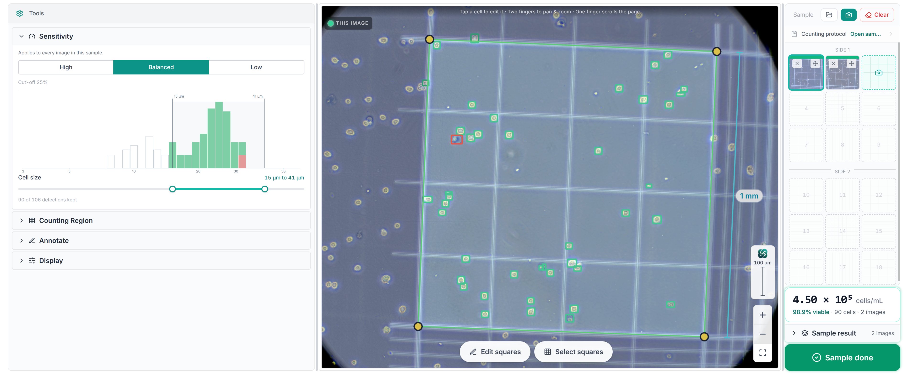
What comes back
- Per image
Live and dead counts, with every detected cell marked on the image
- Concentration
Cells per mL, from the grid measured in the image and the dilution
- Viability
The live share of counted cells, in percent
- Size
A cell-size histogram pooled over the sample, with the gate drawn on it
- Corrections
Add a missed cell, delete a wrong one, flip live to dead, re-run at another sensitivity
Nothing is final until the person who knows the sample accepts the count.
Results come back in seconds for a typical frame, and in under 30 seconds for a twelve-megapixel image. The accepted count is dated and names who accepted it, and leaves as a results document or an export bundle. Everything that comes back.
How it was tested
On 16 images taken straight from a leading automated cell counter, none of which CellCount had seen, concordance on cell concentration was 0.91 (Lin’s coefficient), and the live-or-dead call agreed on 95% of the 6,765 cells both readers found (benchmark report of ). On held-out frames the shipped model had never seen, its live-or-dead call agreed with an expert annotator on 98.7% of the 2,682 cells both found.
The method, the data and every number are kept with the study and are available on request. The benchmark, with its plots.
What it counts on
- Resolution
The camera’s full resolution
Frames under about 2,000 px on the short side are too small to count reliably. Images are uploaded as captured: nothing is resized on the way in, because resizing moves the count and compression does not.
- Chamber
Improved Neubauer hemocytometer, bright-line ruling
One full 1 mm square, with all four corners, in the frame. The rulings in the image are what the scale is measured from.
- Stain
Trypan blue
Clear cells are counted live, blue cells dead.
The word is also spelled haemocytometer, in British English, or hemacytometer, and the device is also called a counting chamber or a Neubauer chamber. CellCount is a general-purpose cell detector with viability: it does not classify cell types, and the cell-size gate leaves out what is too small or too large for your cells. The image requirements in full.
Short answers
Is there an app that counts hemocytometer cells automatically?
Yes. CellCount is a web app that counts the cells in a photo of a hemocytometer. It finds the grid in the image, counts the live and dead cells in the squares your protocol names, and works out cells per mL from the measured grid and the dilution you set. If the rulings cannot be found, you place the crossings by hand. It runs in the browser on a laptop, a tablet or a phone, with nothing to install.
Do I draw the squares?
No. CellCount finds the grid by itself: the rulings give it the scale and the square to count in, and the volume follows from them. There are no regions to draw and nothing to calibrate first. If the rulings cannot be found, in a blurred photo for example, you place the crossings by hand.
Does it work from a phone?
Yes. Hold a modern phone to the eyepiece, focus on the rulings and photograph one full 1 mm square with all four corners in the frame, at full resolution. No adapter and no hardware. For cells of about ten micrometres and larger a phone photo counts well; small cells such as yeast want a camera on the microscope port.
Is it free?
Yes, on the Free plan, with no card needed: sign up and count the same day with a phone at the eyepiece or a camera on the microscope. On the Free plan, counted images may be used to improve the service, including our models. Labs that need more get a plan shaped around them.